Jellyfish-inspired device that rapidly and efficiently captures cancer cells from blood samples could enable better patient monitoring

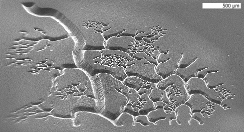
Cells traveling through a microfluidic device can be trapped
by strands of DNA (green).
Image: Suman Bose and Chong Shen

Tumor cells circulating in a patient’s bloodstream can yield a great deal of information on how a tumor is responding to treatment and what drugs might be more effective against it. But first, these rare cells have to be captured and isolated from the many other cells found in a blood sample.

Many scientists are now working on microfluidic devices that can isolate circulating tumor cells (CTCs), but most of these have two major limitations: It takes too long to process a sufficient amount of blood, and there is no good way to extract cancer cells for analysis after their capture.

A new device from researchers at MIT and Brigham and Women’s Hospital overcomes those obstacles. Inspired by the tentacles of a jellyfish, the team coated a microfluidic channel with long strands of DNA that grab specific proteins found on the surfaces of leukemia cells as they flow by. Using this strategy, the researchers achieved flow rates 10 times higher than existing devices — fast enough to make the systems practical for clinical use.

Using this technology, described in this week’s issue of the Proceedings of the National Academy of Sciences, doctors could monitor cancer patients to determine whether their treatment is working.

“If you had a rapid test that could tell you whether there are more or less of these cells over time, that would help to monitor the progression of therapy and progression of the disease,” says Jeff Karp, an associate professor of medicine at Harvard Medical School and co-director of the Center for Regenerative Therapeutics at Brigham and Women’s Hospital in Boston.

This type of device could also enable personalized treatments: Once cells are isolated from a patient, doctors could test different drugs on them to determine which are most effective.

DNA ‘tentacles’

The number of CTCs found in a milliliter of a particular patient’s blood can range from just a few to several thousand. To isolate those rare cells, researchers have tried building microfluidic channels dotted with antibodies specific to a protein found on the target cells. However, because the antibodies only extend tens of nanometers from the bottom of the channel, the capture of cells by the antibodies is slow.

To extend the reach of the capture molecules, Karp and Karnik’s team mimicked the tentacles of jellyfish, creating long strands of repeating DNA sequences. Those sequences, known as aptamers, target a protein found in large numbers on leukemia cells.

The DNA strands are attached to a microchannel with a herringbone pattern on its floor. Those patterned ridges cause the blood to swirl as it flows through the channel, improving the chances that individual cells will come into contact with the tentacles, which extend hundreds of microns into the channel. This allows the researchers to increase the rate of blood flow.

“Normally what happens at high flow rates is the cells don’t really come close to the surface, and it’s very challenging to capture the target cells,” Karnik says. “But this combination of these herringbone grooves to mix the solution and bring the cells into contact with surfaces, plus having aptamers that are sticking out into the solution, enables very high capture rates at very high flow rates.”

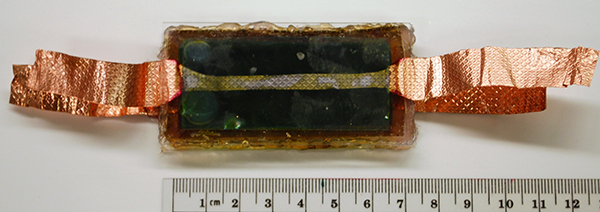
Flow rates in the new device are 10 times higher than those reported for previous devices, and the system can capture 60 to 80 percent of the target cells. In the current model, which measures 1 square centimeter, the flow rate is 1 milliliter per hour. By making the device larger, the researchers say they could boost the flow rate to 100 milliliters of blood per hour — fast enough to rapidly process the 10- to 20-milliliter samples that would be needed to get an accurate CTC count from an individual patient.

Because the “tentacles” are made of DNA, they can easily be cleaved with enzymes, freeing the captured cells for further analysis.

Peter Kuhn, an associate professor of cell biology at the Scripps Research Institute, says this technology is an interesting alternative to existing approaches to capture rare cells, adding that further testing is needed to validate the technology’s usefulness.

“This field needs many different avenues for rare-cell identification to be explored,” Kuhn says. “These technologies can provide the key enablers for studying disease in humans.”

Minimally invasive

Devices that capture CTCs could offer a better alternative to sampling bone marrow in determining whether cancer treatment is working in a leukemia patient.

“If one could improve the sensitivity of detection in blood, then this approach may enable a transition from isolating marrow to isolating blood, which is much less invasive and you can do it more often. It could change the paradigm for how residual disease is detected,” Karp says.

“The beauty of this technology is its versatility,” Zhao says. “You can easily modify the length and density of the DNA chains; you can include different sequences in the DNA to capture different types of cells.”

This feature makes it a platform technology that can be broadly applied in the clinic and research laboratories. For instance, another possible application is capturing fetal cells, which are very rare in a pregnant woman’s bloodstream. Analyzing these cells could help doctors perform prenatal diagnostic tests for a range of diseases using an approach that is far less invasive than amniocentesis.

The researchers are now working on adapting the DNA strands to target other molecules, such as receptors found on the surfaces of cells dislodged from solid tumors
source:http://web.mit.edu/newsoffice/2012/capturing-rare-cancer-cells-1112.html

Nanoengineers can print 3D microstructures in mere seconds

NanoEngineering Professor Shaochen Chen has demonstrated the capability of printing three-dimensional blood vessels in mere seconds out of soft, biocompatible hydrogels. Being able to print blood vessels is essential to achieving the promise of regenerative medicine because it is how the body distributes oxygen and nutrients. Image Credit: Biomedical Nanotechnology Laboratory, Chen Research Group, UC San Diego Jacobs School of Engineering. All rights reserved by Jacobs School of Engineering

Nanoengineers at the University of California, San Diego have developed a novel technology that can fabricate, in mere seconds, microscale three dimensional (3D) structures out of soft, biocompatible hydrogels. Near term, the technology could lead to better systems for growing and studying cells, including stem cells, in the laboratory. Long-term, the goal is to be able to print biological tissues for regenerative medicine. For example, in the future, doctors may repair the damage caused by heart attack by replacing it with tissue that rolled off of a printer.
Reported in the journal Advanced Materials, the biofabrication technology, called dynamic optical projection stereolithography (DOPsL), was developed in the laboratory of NanoEngineering Professor Shaochen Chen. Current fabrication techniques, such as photolithography and micro-contact printing, are limited to generating simple geometries or 2D patterns. Stereolithography is best known for its ability to print large objects such as tools and car parts. The difference, says Chen, is in the micro- and nanoscale resolution required to print tissues that mimic nature’s fine-grained details, including blood vessels, which are essential for distributing nutrients and oxygen throughout the body. Without the ability to print vasculature, an engineered liver or kidney, for example, is useless in regenerative medicine. With DOPsL, Chen’s team was able to achieve more complex geometries common in nature such as flowers, spirals and hemispheres.  Other current 3D fabrication techniques, such as two-photon photopolymerization, can take hours to fabricate a 3D part.
The biofabrication technique uses a computer projection system and precisely controlled micromirrors to shine light on a selected area of a solution containing photo-sensitive biopolymers and cells. This photo-induced solidification process forms one layer of solid structure at a time, but in a continuous fashion.
The Chen Research Group is focused on fabrication of nanostructured biomaterials and nanophotonics for biomedical engineering applications
source: http://www.jacobsschool.ucsd.edu/news/news_releases/release.sfe?id=1259

Spinach power gets a major boost

An interdisciplinary team of researchers at Vanderbilt University have developed a way to combine the photosynthetic protein that converts light into electrochemical energy in spinach with silicon, the material used in solar cells, in a fashion that produces substantially more electrical current than has been reported by previous “biohybrid” solar cells.
“This combination produces current levels almost 1,000 times higher than we were able to achieve by depositing the protein on various types of metals. It also produces a modest increase in voltage,” said David Cliffel, associate professor of chemistry, who collaborated on the project with Kane Jennings, professor of chemical and biomolecular engineering.
“If we can continue on our current trajectory of increasing voltage and current levels, we could reach the range of mature solar conversion technologies in three years.”
The researchers’ next step is to build a functioning PS1-silicon solar cell using this new design. Jennings has an Environmental Protection Agency award that will allow a group of undergraduate engineering students to build the prototype. The students won the award at the National Sustainable Design Expo in April based on a solar panel that they had created using a two-year old design. With the new design, Jennings estimates that a two-foot panel could put out at least 100 milliamps at one volt – enough to power a number of different types of small electrical devices.

Harnessing the power of spinach

More than 40 years ago, scientists discovered that one of the proteins involved in photosynthesis, called Photosystem 1 (PS1), continued to function when it was extracted from plants like spinach. Then they determined PS1 converts sunlight into electrical energy with nearly 100 percent efficiency, compared to conversion efficiencies of less than 40 percent achieved by manmade devices. This prompted various research groups around the world to begin trying to use PS1 to create more efficient solar cell

Another potential advantage of these biohybrid cells is that they can be made from cheap and readily available materials, unlike many microelectronic devices that require rare and expensive materials like platinum or indium. Most plants use the same photosynthetic proteins as spinach. In fact, in another research project Jennings is working on a method for extracting PS1 from kudzu.
Since the initial discovery, progress has been slow but steady. Researchers have developed ways to extract PS1 efficiently from leaves. They have demonstrated that it can be made into cells that produce electrical current when exposed to sunlight. However, the amount of power that these biohybrid cells can produce per square inch has been substantially below that of commercial photovoltaic cells.
Another problem has been longevity. The performance of some early test cells deteriorated after only a few weeks. In 2010, however, the Vanderbilt team kept a PS1 cell working for nine months with no deterioration in performance.

“Nature knows how to do this extremely well. In evergreen trees, for example, PS1 lasts for years,” said Cliffel. “We just have to figure out how to do it ourselves.”

Secret is “doping” silicon

The Vanderbilt researchers report that their PS1/silicon combination produces nearly a milliamp (850 microamps) of current per square centimeter at 0.3 volts. That is nearly two and a half times more current than the best level reported previously from a biohybrid cell. The reason this combo works so well is because the electrical properties of the silicon substrate have been tailored to fit those of the PS1 molecule. This is done by
implanting electrically charge atoms in the silicon to alter its electrical properties: a process called “doping.” In this case, the protein worked extremely well with silicon doped with positive charges and worked poorly with negatively doped silicon.
To make the device, the researchers extracted PS1 from spinach into an aqueous solution and poured the mixture on the surface of a p-doped silicon wafer. Then they put the wafer in a vacuum chamber in order to evaporate the water away leaving a film of protein. They found that the optimum thickness was about one micron, about 100 PS1 molecules thick.

Protein alignment

When a PS1 protein exposed to light, it absorbs the energy in the photons and uses it to free electrons and transport them to one side of the protein. That creates regions of positive charge, called holes, which move to the opposite side of the protein.
In a leaf, all the PS1 proteins are aligned. But in the protein layer on the device, individual proteins are oriented randomly. Previous modeling work indicated that this was a major problem. When the proteins are deposited on a metallic substrate, those that are oriented in one direction provide electrons that the metal collects
while those that are oriented in the opposite direction pull electrons out of the metal in order to fill the holes that they produce. As a result, they produce both positive and negative currents that cancel each other out to leave a very small net current flow.
The p-doped silicon eliminates this problem because it allows electrons to flow into PS1 but will not accept them from protein. In this manner, electrons flow through the circuit in a common direction.

source : http://news.vanderbilt.edu/2012/09/spinach-power-a-major-boost/photo-2-241_20120828170545-biohybridcell600/

The Laser Beam as a “3D Painter”

3D pattern, produced by photografting (180 µm wide). Fluorescent molecules are attached to the hydrogel, resulting in a microscopic 3D pattern.


There are many ways to create three dimensional objects on a micrometer scale. But how can the chemical properties of a material be tuned at micrometer  precision? Scientists at the Vienna University of Technology developed a method to attach molecules at exactly the right place. When biological tissue is grown, this method can allow the positioning of chemical signals, telling living cells where to attach. The new technique also holds promise for sensor technology: A tiny three dimensional “lab on a chip” could be created, in which accurately positioned molecules react with substances from the environment.

Materials Science and Chemistry
“3D-photografting” is the name of the new method. Two research teams from the Vienna University of Technology collaborated closely to develop it: Professor Jürgen Stampfl’s materials science team and Professor Robert Liska’s research group for macromolecular chemistry.

Both research groups have already attracted considerable attention in the past, developing new kinds of 3D-printers. However, for the applications on which the scientists are working on now, 3D-printing would not have been useful: “Putting together a material from tiny building blocks with different chemical properties would be extremely complicated”, says Aleksandr Ovsianikov. “That is why we start from a three dimensional scaffold and then attach the desired molecules at exactly the right positions.”

Molecules in the Hydrogel – Locked into Position by the Laser
The scientists start with a so-called hydrogel – a material made of macromolecules, arranged in a loose meshwork. Between those molecules, large pores remain, through which other molecules or even cells can migrate.
Specially selected molecules are introduced into the hydrogel meshwork, then certain points are irradiated with a laser beam. At the positions where the focused laser beam is most intense, a photochemically labile bond is broken. That way, highly reactive intermediates are created which locally attach to the hydrogel very quickly. The precision depends on the laser’s lens system, at the Vienna University of Technology a resolution of 4 µm could be obtained. “Much like an artist, placing colors at certain points of the canvas, we can place molecules in the hydrogel – but in three dimensions and with high precision”, says Aleksandr Ovsianikov.

Chemical Signals for Cells
This method can be used to artificially grow biological tissue. Like a climbing plant clinging to a rack, cells need some scaffold at which they attach. In a natural tissue, the extracellular matrix does the trick by using specific amino acid sequences to signal the cells, where they are supposed to grow.
In the lab, scientists are trying to use similar chemical signals. In various experiments, cell  attachment could be guided on two dimensional surfaces, but in order to grow larger tissues with a specific inner structure (such as capillaries), a truly three dimensional technique is required.

Micro Sensors Detect Molecules
Depending on the application, different molecules can be used. 3D photografting is not only useful for bio-engineering but also for other fields, such as photovoltaics or sensor technology. In a very small space, molecules can be positioned which attach to specific chemical substances and allow their detection. A microscopic three-dimensional “lab on a chip” becomes possible.

source: http://www.tuwien.ac.at/en/news/news_detail/article/7719/

Mathematicians Solve Decade-Old Debate On Regulation of Protein Production by microRNAs in Cells

An international team of mathematicians has proposed a new solution to understanding a biological puzzle that has confounded molecular biologists

This is an illustration of the model applied by Professor Gorban.  

They have applied a mathematical model to work out the functioning of small molecules known as microRNAs — components of the body akin to the electronics in modern airplanes.
For a long time molecular biologists thought that the major role of RNA in living cells was to serve as a copy of a gene and a template for producing proteins, major cell building blocks. This belief had been changed at the end of 90s when it was found that myriads of RNA molecules are involved in regulating speeds of practically all molecular mechanisms in a cell. These abundant molecules are essential in regulating the speed of protein production- a vital function in bodily processes, including development, differentiation and cancer.
The problem to date has been that scientists have differed over interpretations of how the production of the major building blocks of a cell, proteins, is controlled by microRNAs.
Basically, there were different and sometimes conflicting theories about ways in which microRNAs regulate protein production since the results varied depending on only slightly changed experimental conditions.
Professor Alexander Gorban, who holds a Chair in Applied Mathematics at University of Leicester, said: “The old metaphor of an elephant and blind scientists trying to describe it will be always relevant to science. However, often we use it only as a metaphor, as a generic statement. In this project the elephant’s metaphor can be applied literally as a working principle.
“Different biological labs or slightly changed experimental conditions meant that results were different for investigators.
“Quite dramatically, there has been a series of reports in top-ranked journals with contradictory results supporting one or another mechanism. Furthermore, researchers are puzzled by the fact that the same couple of protein and microRNA demonstrate different mechanisms of regulation in different biological labs or in slightly changed experimental conditions.”
The mathematical model constructed by Professor Gorban from University of Leicester and Andrei Zinovyev from Institut Curie in Paris in collaboration with biologists Nadya Morozova and Annick Harel-Bellan from CNRS in France showed that there might be one simple mechanism which manifests itself differently in different conditions. Their findings are due to be published in the RNA Journal.
Professor Gorban said: “We have shown that what appeared to be very different mechanisms are in fact manifestations of one relatively simple biochemical reaction, but taking place in various contexts.
“Our model proposes that microRNA performs many actions simultaneously to the protein development, basically acting to get the job done (regulating the speed of protein production) in a stable and efficient way, given whatever conditions the experiment is occurring in.
“If this model is accepted, we would be able to take active steps in determining what the main mechanism of microRNA action is, as the model suggests experiments to verify the hypothesis. This in turn should lead to a resolution of a decade long debate to understand the means in which these very important molecules actually work.”
Pat Heslop-Harrison, Professor of Cell Biology at the University of Leicester, said: “The discovery of miRNA and its regulatory role has completely changed our view of how genes in cells are controlled.
“Understanding all the ways the regulation is happening and interpreting experimental evidence has proved a huge challenge. In this important new paper, Alexander, Andrei, Nadya and colleagues overview the characteristic features of no less than nine different mechanisms, and then generate a unifying model of the whole system integrating the nine mechanisms.
“The multifunctional model gives dynamic predictions of gene control; it can now be tested to understand significance of the various mechanisms coexisiting under different conditions. It will be exciting to link this back the huge range of functions and responses of organisms and understanding miRNA control mechanisms is a systematic and predictive way.”

From Microns to Centimeters: Researchers Invent New Tissue Engineering Tool

Image of cells aligned to spell “Toronto.” (Credit: courtesy Lian Leng)

Along with graduate students from their labs — Lian Leng, Boyang Zhang, and Arianna McAllister — Associate Professor Axel Guenther of the Department of Mechanical and Industrial Engineering, cross-appointed to the Institute of Biomaterials and Biomedical Engineering (IBBME), and Associate Professor Milica Radisic, core professor at IBBME and the Department of Chemical Engineering and Applied Chemistry, have invented a new device that may allow for the uniform, large-scale engineering of tissue.
“There’s a lot of interest in soft materials, particularly biomaterials,” explains Guenther of the materials that help create functional tissue cultures, “but until now no one has demonstrated a simple and scalable one-step process to go from microns to centimeters.”
The invention, presented in a cover article for the journal Advanced Materials this month, is currently being commercialized by MaRS Innovations in collaboration with the Innovations and Partnerships Office (IPO) of the University of Toronto, where Radisic and Guenther’s labs have filed two patents on the device.
But how exactly does a machine grow a large patch of living tissue?
Scientists manipulate biomaterials into the micro-device through several channels. The biomaterials are then mixed, causing a chemical reaction that forms a “mosaic hydrogel” — a sheet-like substance compatible with the growth of cells into living tissues, into which different types of cells can be seeded in very precise and controlled placements, according to the researchers.
Unique to this new approach to tissue engineering, however, and unlike more typical methods for tissue engineering (for instance, scaffolding, the seeding of cells onto an artificial structure capable of supporting three-dimensional tissue formation) cells planted onto the mosaic hydrogel sheets are precisely incorporated into the mosaic hydrogel sheet just at the time it’s being created — generating the perfect conditions for cells to grow.
The placement of the cells is so precise, in fact, that scientists can spell words (such as “Toronto,” shown here) and can precisely mimic the natural placement of cells in living tissues. And by collecting these sheets around a drum, the machine is able to collect layers of cells in thicknesses made to measure: in essence, three dimensional, functional tissues.
And in tissue engineering, cell placement is everything: something that the new invention delivers. “The cells are able to stretch and connect with each other, which is very important for ultimately obtaining functional tissues,” Guenther states.
The resulting tissues, cites Lian Leng, lead author on the project and a 3rd year PhD Candidate in the Department of Mechanical and Industrial Engineering, are remarkably stable. “In this case, when we put the cells in the right places we create cellular organization quite naturally.”
“My laboratory is currently pursuing different applications of the technology — different tissues,” says Guenther. The device may provide the means to create three-dimensional cell cultures for the development of therapeutic drugs, for instance. “But one of my dreams is to one day engineer a vascularized leaf — perhaps a maple leaf,” he jokes.

Scientists Create First Computerized Model Of Organism’s Entire Lifecycle

For the first time, researchers at Stanford University in California have used computer software to simulate the entire lifecycle of an organism — all 525 genes of the bacterium Mycoplasma genitalium.
The breakthrough, which is described in a paper published in the journal Cell on July 20, could lead to enormous advances in the fields of genetics, personalized medicine, pharmaceuticals and biology in general, allowing scientists to perform experiments that wouldn’t be possible on actual living organisms. Researchers used 128 computers to model an organism that is actually a sexually transmitted parasite.
“I’m a big believer in ‘model-driven discovery’ which simply means that if you have a model of a biological process, you will make discoveries more quickly and efficiently than you would without a model,” said Markus Covert, an assistant professor of bioengineering at Stanford, who led the study, in an email to TPM.
Specifically, Covert explained Stanford’s software simulation of the Mycoplasma genitalium lifecycle is distinct from previous efforts to model organisms using computers because of the vast volume of the data it encompasses — literally every single molecular process that takes place in the single-celled bacterium’s life.
Covert’s team has already begun experimenting with removing certain genes to see what happens in the simulation.
“Each simulation is one cell, one life cycle,” Covert said. “We’ve now run thousands of these, both for the normally growing cell as well as all of the single-gene knockout strains.”
The simulation takes about nine to 10 hours in order for one cell to divide, again emulating its real counterpart, as The New York Times reported about the study. The simulation stops when the organism “dies.”
“‘Death’ of the simulation is defined as an inability to divide, degradation of key components, etc,” Covert explained.
The Standford simulation of the parasite has already led to one important discovery: The finding that the longer it takes for a single cell to begin replicating its DNA in preparation for division, the shorter time it will actually take to replicate, which balances out to the same average time across all cells modeled.
Among the many new advances that the computer simulation could lead to is the possibility “for the wholesale creation of new microorganisms,” according to a Stanford news release on the work.
Still, it was a long road to get from the initial idea to simulate the bacterium’s life cycle on a computer to actually doing it, as covert explained.
“I first thought about getting involved in whole-cell modeling about thirteen years ago, when I read a quote by a scientist in the paper who said (paraphrasing) that the ultimate challenge in biology would be to create a computer model of a cell, because it would imply a fundamental understanding of how cells work,” Covert wrote. “I basically became obsessed with that quote, and still think about it every day!”
It took nearly four years to actually write the software to perform the simulation itself, and the use of over 900 prior scientific papers on Mycoplasma genitalium and other bacterium to create the perfect algorithm to simulate the organism. The research was funded in part by the National Institutes of Health.

Bacterium engineered with DNA in which thymine is replaced by synthetic building block

Source: Bacterium engineered with DNA in which thymine is replaced by synthetic building block

The project, coordinated by Rupert Mutzel (Institut für Biologie, Freie Universität Berlin) and Philippe Marlière (Heurisko USA Inc.), involved researchers of the French CEA (Commissariat à l’Energie Atomique et aux Energies Alternatives) and of the Katholieke Universiteit Leuven (Belgium). As described in the latest issue ofAngewandte Chemie International Edition, the experimental work was based on a unique technology developed by Marlière and Mutzel enabling the directed evolution of organisms under strictly controlled conditions. Large populations of microbial cells are cultured for prolonged periods in the presence of a toxic chemical — in this case, 5-chlorouracil — at sublethal levels, thereby selecting for genetic variants capable of tolerating higher concentrations of the toxic substance.
In response to the appearance of such variants in the cell population the concentration of the toxic chemical in the growth medium is increased, thus keeping the selection pressure constant. This automated procedure of long term evolution was applied to adapt genetically engineered Escherichia colibacteria unable to synthesize the natural nucleobase thymine to grow on increasing concentrations of 5-chlorouracil. After a culture period of about 1000 generations descendants of the original strain were obtained which used 5-chlorouracil as complete substitute for thymine. Subsequent genome analysis revealed numerous mutations in the DNA of the adapted bacteria. The contribution of these mutations to the adaptation of the cells towards the halogenated base will be the subject of follow-up studies.
Besides the obvious interest of this radical change in the chemistry of living systems for basic research the scientists consider the outcome of their work also to be of importance for “xenobiology,” a branch of synthetic biology. This young area of the life sciences aims at the generation of new organisms not found in nature harboring metabolic traits optimized for alternative modes of energy production or for the synthesis of high value chemicals. Like GMOs, such organisms are seen as a potential threat for natural ecosystems when released from their laboratory confinements, either through direct competition with wild type organisms or through diffusion of their “synthetic” DNA.
Scientists have recognized that physical containment cannot in every single case prevent engineered live forms from reaching natural habitats, in the same way as radioactive isotopes can leak into the surroundings of a nuclear power plant. However, synthetic organisms like those evolved by Marlière and Mutzel and their collaborators which depend on the availability of substances for their proliferation not found in nature or which incorporate non-natural building blocks in their genetic material could neither compete nor exchange genetic messages with wild type organisms, but would die in the absence of the xenobiotic.

Ants Are Experienced Fungus Farmers

Source: Ants Are Experienced Fungus Farmers

Entomologists Ted Schultz and Seán Brady at the Smithsonian’s National Museum of Natural History have been providing new insight into the agricultural abilities of ants and how these abilities have evolved throughout time. Using DNA sequencing, the scientists were able to construct an “evolutionary tree” of fungus-growing ants, which revealed a single pioneering ancestor that discovered agriculture approximately 50 million years ago.
In the past 25 million years, four different specialized agricultural systems have evolved, leading to the most recently evolved and best-known fungus-growing ant species–“leaf-cutter ants.” The ants do not eat the leaves; they grow their fungus gardens on them and then eat the fungus. By studying the agricultural evolution of leaf-cutter ants, as well as various other species, scientists may be able to develop improved human agricultural and medical methods.
“Agriculture is very rare in the animal world,” said Schultz. “We only know of four animal groups that have discovered agriculture: ants, termites, bark beetles and humans. By studying certain fungus-growing ants, which our study indicates are almost like ‘living fossils,’ we might be able to better understand steps involved in the evolution of ant agriculture.”
To complete their research, scientists spent more than 15 years assembling a comprehensive array of specimens, which included 91 ant species, 65 of which were fungus-growing ant species representing all different groups of fungus-growing ants. Researchers then used DNA sequencing, combined with a variety of state-of-the-art computer algorithms, to construct an evolutionary tree of fungus-growing ants. Dominican amber fungus-growing ant fossils were used to calibrate time intervals on the evolutionary tree.
From this evolutionary tree, scientists were able to determine that fungus-growing ants are all descended from a common ancestor that pioneered agriculture 50 million years ago during a period of global warming. The researchers also determined that in the past 25 million years, four different specialized agricultural systems emerged.
Each of these systems has its own unique set of cultivated fungi. For example, approximately 20 million years ago one group of fungus-growing ants discovered “higher agriculture,” meaning they cultivated their fungi to produce specialized “fruits” that the ants would harvest and eat for food. Leaf-cutter ants, which belong to this group, originated recently–less than 10 million years ago. Finally, it also was discovered that there are certain fungus-growing ant species living in South America today that are “missing links” in evolution.

Nanotechnology used to harness power of fireflies

Source: Nanotechnology used to harness power of fireflies


It’s all about the size and structure of the custom, quantum nanorods, which are produced in the laboratory by Mathew Maye, assistant professor of chemistry in SU’s College of Arts and Sciences; and Rebeka Alam, a chemistry Ph.D. candidate. Maye is also a member of the Syracuse Biomaterials Institute. “Firefly light is one of nature’s best examples of bioluminescence,” Maye says. “The light is extremely bright and efficient. We’ve found a new way to harness biology for non-biological applications by manipulating the interface between the biological and non-biological components.”

Their work, “Designing Quantum Rods for Optimized Energy Transfer with Firefly Luciferase Enzymes,” was published online May 23 in Nano Letters and is forthcoming in print. Collaborating on the research were Professor Bruce Branchini and Danielle Fontaine, both from Connecticut College.
Fireflies produce light through a chemical reaction between luciferin and it’s counterpart, the enzyme luciferase. In Maye’s laboratory, the enzyme is attached to the nanorod’s surface; luciferin, which is added later, serves as the fuel. The energy that is released when the fuel and the enzyme interact is transferred to the nanorods, causing them to glow. The process is called Bioluminescence Resonance Energy Transfer (BRET).
“The trick to increasing the efficiency of the system is to decrease the distance between the enzyme and the surface of the rod and to optimize the rod’s architecture,” Maye says. “We designed a way to chemically attach, genetically manipulated luciferase enzymes directly to the surface of the nanorod.” Maye’s collaborators at Connecticut College provided the genetically manipulated luciferase enzyme.
The nanorods are composed of an outer shell of cadmium sulfide and an inner core of cadmium seleneide. Both are semiconductor metals. Manipulating the size of the core, and the length of the rod, alters the color of the light that is produced. The colors produced in the laboratory are not possible for fireflies. Maye’s nanorods glow green, orange, and red. Fireflies naturally emit a yellowish glow. The efficiency of the system is measured on a BRET scale. The researchers found their most efficient rods (BRET scale of 44) occurred for a special rod architecture (called rod-in-rod) that emitted light in the near-infrared light range. Infrared light has longer wavelengths than visible light and is invisible to the eye. Infrared illumination is important for such things as night vision goggles, telescopes, cameras, and medical imaging.
Maye’s and Alam’s firefly-conjugated nanorods currently exist only in their chemistry laboratory. Additional research is ongoing to develop methods of sustaining the chemical reaction — and energy transfer — for longer periods of time and to “scale-up” the system. Maye believes the system holds the most promise for future technologies that that will convert chemical energy directly to light; however, the idea of glowing nanorods substituting for LED lights is not the stuff of science fiction.